Home » Uncategories » Diagram Of Chest Area / Anatomical Terminology Anatomy And Physiology I - Pectoralis major trigger point diagram, pain patterns and related medical symptoms.
Diagram Of Chest Area / Anatomical Terminology Anatomy And Physiology I - Pectoralis major trigger point diagram, pain patterns and related medical symptoms.
Diagram Of Chest Area / Anatomical Terminology Anatomy And Physiology I - Pectoralis major trigger point diagram, pain patterns and related medical symptoms.. Possible causes of pain include trauma, musculoskeletal. The hindquarters encompass the he large muscular area of the hind legs and are located above the stifle and behind the barrel. Strains and inflammation in the chest wall muscles or ribs usually goes away on its own. This is the area from the bottom end of the neck to the top of the front legs. The heart is enclosed in the pericardium which is a double layer.
It also protects several vital organs of the chest, such as the heart, aorta, vena cava, and thymus gland that are located just deep to the sternum. Profile view of female chest area. In the human body, the region of the thorax between the neck and diaphragm in the front of the body is called the chest. Your heart is surrounded by important blood vessels and arteries which pump blood into and out of your heart. Sensory information from the body and critical signals.
Tight Chest Muscles Why Your Upper Back Is The Key To Their Release Laguna Orthopedic Rehabilitation from images.squarespace-cdn.com This is the area from the bottom end of the neck to the top of the front legs. Strains and inflammation in the chest wall muscles or ribs usually goes away on its own. It lies between the right and left lungs, in the middle of the chest and slightly towards the left of the breastbone. The corresponding area in an animal can also be referred to as the chest. This page provides an overview of the chest muscle group. The heart is enclosed in the pericardium which is a double layer. Profile view of female chest area. Thoracic cavity, also called chest cavity, the second largest hollow space of the body.it is enclosed by the ribs, the vertebral column, and the sternum, or breastbone, and is separated from the abdominal cavity (the body's largest hollow space) by a muscular and membranous partition, the diaphragm.it contains the lungs, the middle and lower airways—the tracheobronchial tree—the heart.
Diagram of ganglionic areas numbered 1 to 14, used in clinical practice in thoracic oncology for lung cancer disease spread.
Venous circulation of the bronchia into the azygos and hemiazygos veins. Location of chest pain during angina or heart attack diagram in this image, you will find an upper chest, substernal radiating to neck and jaw, substernal raiding down left arm, substernal radiating down left arm, epigastric radiating to neck, jaw, and arms, neck and jaw, left shoulder and down both arms, intrascapular in it. Thoracic cavity, also called chest cavity, the second largest hollow space of the body.it is enclosed by the ribs, the vertebral column, and the sternum, or breastbone, and is separated from the abdominal cavity (the body's largest hollow space) by a muscular and membranous partition, the diaphragm.it contains the lungs, the middle and lower airways—the tracheobronchial tree—the heart. Strains and inflammation in the chest wall muscles or ribs usually goes away on its own. Anatomy of machine 12 photos of the anatomy of machine anatomy of a fog machine, anatomy of a. The myofascial pain pattern has pain locations that are displayed in red and associated trigger points shown as xs. Sensory information from the body and critical signals. A woman's chest — like the rest of her body — is covered with skin that has two layers. It is a flat bone about six inches in length, around an inch wide, and only a fraction of an. The corresponding area in an animal can also be referred to as the chest. Powerful muscles that move the head and arms attach to these bones as well. The epidermis is the outermost layer that provides a protective, waterproof seal over the body. It also has branching energy pathways all over the chest.
The hindquarters encompass the he large muscular area of the hind legs and are located above the stifle and behind the barrel. The chest wall thoracic lymph nodes receive drainage from the breasts, arms, pectoral muscles, and other muscles and skin located in the upper section of the chest. Profile view of female chest area. Related posts of anatomy of the chest area anatomy of machine. This is the area from the bottom end of the neck to the top of the front legs.
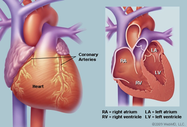
Human Heart Anatomy Diagram Function Chambers Location In Body from img.webmd.com The anatomy of the human ribs (costae) are one of the integral parts of the chest wall; Sensory information from the body and critical signals. The circulatory system does most of its. It lies between the right and left lungs, in the middle of the chest and slightly towards the left of the breastbone. The hindquarters encompass the he large muscular area of the hind legs and are located above the stifle and behind the barrel. Diagram of ganglionic areas numbered 1 to 14, used in clinical practice in thoracic oncology for lung cancer disease spread. This pericardium is attached to the diaphragm, spinal column and other parts via strong ligaments. Last medically reviewed on july.
The chest is the area of origin for many of the body's systems as it houses organs such as the heart, esophagus, trachea, lungs, and thoracic diaphragm.
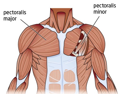
Pectoralis major trigger point diagram, pain patterns and related medical symptoms. Man head and chest anatomy diagram with ghost effect. The major muscle in the chest is the pectoralis major. Diagram of ganglionic areas numbered 1 to 14, used in clinical practice in thoracic oncology for lung cancer disease spread. This page provides an overview of the chest muscle group. Location of chest pain during angina or heart attack diagram in this image, you will find an upper chest, substernal radiating to neck and jaw, substernal raiding down left arm, substernal radiating down left arm, epigastric radiating to neck, jaw, and arms, neck and jaw, left shoulder and down both arms, intrascapular in it. The anatomy of the human ribs (costae) are one of the integral parts of the chest wall; The epidermis is the outermost layer that provides a protective, waterproof seal over the body. Sensory information from the body and critical signals. That's all of the acupuncture points that are a located on the chest area of the human body. Find out more about the individual muscles within the chest anatomy by clicking their respective links throughout this It also protects several vital organs of the chest, such as the heart, aorta, vena cava, and thymus gland that are located just deep to the sternum. Anatomy of machine 12 photos of the anatomy of machine anatomy of a fog machine, anatomy of a.
The diaphragm, a sheet of muscle in the middle chest area, is essential for breathing. That's all of the acupuncture points that are a located on the chest area of the human body. This page provides an overview of the chest muscle group. Connective tissue called the mesentery holds the abdominal organs together. This is the area from the bottom end of the neck to the top of the front legs.
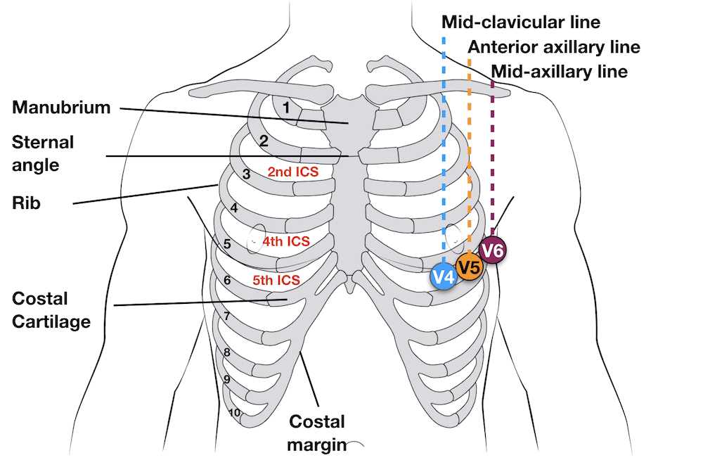
Ecg Lead Positioning Litfl Ecg Library Basics from litfl.com They make up the lateral part of our body, its anterior and posterior wall and they entirely build the lateral parts of the chest wall. The epidermis is the outermost layer that provides a protective, waterproof seal over the body. A woman's chest — like the rest of her body — is covered with skin that has two layers. Any diaphragm pain can, therefore, be very alarming. This is the area from the bottom end of the neck to the top of the front legs. Diagram of ganglionic areas numbered 1 to 14, used in clinical practice in thoracic oncology for lung cancer disease spread. Thoracic cavity, also called chest cavity, the second largest hollow space of the body.it is enclosed by the ribs, the vertebral column, and the sternum, or breastbone, and is separated from the abdominal cavity (the body's largest hollow space) by a muscular and membranous partition, the diaphragm.it contains the lungs, the middle and lower airways—the tracheobronchial tree—the heart. It also protects several vital organs of the chest, such as the heart, aorta, vena cava, and thymus gland that are located just deep to the sternum.
Costochondritis, sometimes called costosternal syndrome or anterior chest wall syndrome, merely indicates pain and tenderness in the costochondral junction, which is the area along the sides of the breastbone where the ribs attach.
It lies between the right and left lungs, in the middle of the chest and slightly towards the left of the breastbone. That's all of the acupuncture points that are a located on the chest area of the human body. Powerful muscles that move the head and arms attach to these bones as well. Thoracic cavity, also called chest cavity, the second largest hollow space of the body.it is enclosed by the ribs, the vertebral column, and the sternum, or breastbone, and is separated from the abdominal cavity (the body's largest hollow space) by a muscular and membranous partition, the diaphragm.it contains the lungs, the middle and lower airways—the tracheobronchial tree—the heart. It also protects several vital organs of the chest, such as the heart, aorta, vena cava, and thymus gland that are located just deep to the sternum. They make up the lateral part of our body, its anterior and posterior wall and they entirely build the lateral parts of the chest wall. Diagram of the emergence of the bronchial arteries in the descending thoracic aorta. Location of chest pain during angina or heart attack diagram in this image, you will find an upper chest, substernal radiating to neck and jaw, substernal raiding down left arm, substernal radiating down left arm, epigastric radiating to neck, jaw, and arms, neck and jaw, left shoulder and down both arms, intrascapular in it. The epidermis is the outermost layer that provides a protective, waterproof seal over the body. It is a flat bone about six inches in length, around an inch wide, and only a fraction of an. Find out more about the individual muscles within the chest anatomy by clicking their respective links throughout this The rib cage also anchors the bones of the head, neck, shoulders, and arms to the trunk of the body. The anatomy of the human ribs (costae) are one of the integral parts of the chest wall;
0 Response to "Diagram Of Chest Area / Anatomical Terminology Anatomy And Physiology I - Pectoralis major trigger point diagram, pain patterns and related medical symptoms."

0 Response to "Diagram Of Chest Area / Anatomical Terminology Anatomy And Physiology I - Pectoralis major trigger point diagram, pain patterns and related medical symptoms."
Post a Comment